Hip Dysplasia Pain in Dogs Pain Meds Reviews
Introduction
Dogs and humans take adult from a mutual ancestor. Both species are vertebrates and terrestrial mammals, with a very similar homologous musculoskeletal structure (Effigy one). Considering of this resemblance in torso structure, sure diseases in both species have a common ground. One of these diseases is hip dysplasia (Hard disk drive). Hard disk drive was showtime described in dogs in the 1930'southward (i) and in humans as early on equally Hippocrates (2). Hd is amend known as canine hip dysplasia (CHD) in dogs, and developmental dysplasia of the hip (DDH) in humans. The prevalence of Hard disk drive in humans varies between 0.1and 10%, depending on the population and definition (iii, 4). In dogs, the prevalence varies betwixt 0 and 73.4%, depending on the breed (five–8).
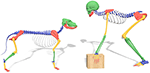
Figure 1. Comparison skeletal construction betwixt dog and human.
There are similar characteristics for Hard disk drive in humans and dogs. In both species the acetabular cover of the femoral caput is insufficient, either because the acetabulum (five, 9, ten) or the femoral head (five, 9, 10) is deformed, or joint laxity (ii, 5) is present. This disturbed femuro-acetabular relationship causes abnormally high peak forces (1, 6, ten) with or without articulation instability and (sub)luxation (two, v, nine) resulting in osteoarthritic changes (2, 5, ix). The torso tries to counter the sequela of Hard disk in both species by thickening and stiffening of the joint capsule (10–12) in order to reduce the laxity (eleven, 12). Still, HD will eventually induce osteoarthritis (OA) resulting in pain (6, 13), lameness (14), and loss of limb function (half-dozen, 13), reducing quality of life.
While CHD and DDH show numerous similar characteristics, disease management is not always the same for both species. In this review nosotros give an overview of the anatomy, etiology, development, diagnostics and handling of HD in humans and dogs. This will provide veterinarians and physicians a perspective and incentive to share the combined translational knowledge.
Anatomy
Initially, the anatomy of humans and dogs may seem very different. For instance, an obvious divergence between dogs and humans is that dogs take a quadruped (4-legged) gait while humans have adopted a bipedal (two-legged) gait. While some anatomical differences have developed due to this difference in gait, dogs and humans have more in common than i might think (Figure one).
The human biped gait has a smaller base of support (less indicate of ground contact) and an elevated center of mass (xv, 16). To balance the body, humans have developed a lumbar lordosis so the center of mass (head, artillery and trunk) is directly above the point of footing contact. This is too more than energy efficient (17, 18). Similarly, a wider pelvis with more laterally oriented iliac crests (as opposed to the coronal airplane in dogs) allowed for some changes in musculature, improving balance on one leg, energy efficiency, and increasing stride length (xvi–18).
While the load orientation (nineteen) of the hip is very similar in dogs and humans, the deviation between biped and quadruped gait gives different load distribution between limbs. Humans distribute their bodyweight betwixt 2 legs while dogs distribute their weight over iv legs, with the front legs carrying approximately 60–65% of the bodyweight (xx–22). Because of the dominance of the front legs over the hind legs, dogs are capable of compensating for hip abnormalities (e.g., HD) by lowering their cervix and increasing the load on the non-affected side (20). Humans can also reduce load on the affected sides by using instruments such as a cane or a stroller (23).
The canine and human anatomy is not just similar on a macroscopic level. Human being and canine hips have a similar cortical microstructure (24, 25) and long os vascularization (25, 26). Because of these external and internal similarities, the dog has long since been (one of) the animal(s) of selection for orthopedic research aimed at humans (24, 27, 28).
Etiology and Pathogenesis
While the verbal etiology of HD for both humans and dogs remains unknown (10, 29), the full general understanding is that both genetic and ecology factors influence the evolution of CHD and DDH (four, 8). First the genetic factors are discussed, followed past ecology factors and finished with the pathogenesis.
Some examples of common genetic factors that influence the occurrence of Hard disk in both species include breed (one, 6) ethnicity (29), increased anteversion angle of the femur (ii, 30, 31), cervix shaft bending of the femur (2, 31), and collagen composition (1, 4). Because of these high genetic factors, family anamnesis is important for discovering Hard disk in humans and improving breeding programs in dogs. Nonetheless, not all genetic factors have an known influence on both species, eastward.g., a articulate genetic factor such equally female sexual activity in humans is known for a college incidence of Hard disk drive (four:1, Female:Male person) (3, 9), while no such relation is known for dogs (8, 32).
Likewise genetic factors there are many different environmental factors influencing the evolution and incidence of HD. Some common environmental factors concern the nutritional state such as nutrition (33), obesity (14, 34, 35) and high nascence weight (5, 36). Furthermore, environmental factors such every bit to seasonal influence (4, 7) and hormone levels have an association with HD (4, 37). Other environmental factors concern disturbances of the biomechanical equilibrium in the pelvic area, e.m., transitional vertebrae (38) tin can change the forces flowing through the hip. This too happens with restrictive swaddling of babies which is common in certain homo cultures. Swaddling limits the abduction and therefore reduces the required strength on the triradiate growth plate (39, xl).
Most of previous mentioned factors are identical between both species, still factors surrounding birth practice differ between species. Humans have but one babe at a time, while dogs have several pups in their litter, meaning intra-uterine mechanical factors are unlike. For case, breech presentation in humans is associated with a loftier incidence of DDH in single child pregnancies, but no relation has been establish in twin pregnancies (3, 41). Similarly, other factors like oligohydramnios (42), breach position, existence showtime built-in (3, iv) and even the preference for the left hip are commonly described in humans, but not in dogs who are typically born in a litter. The preference for the left hip in humans might be explained every bit the left hip is often positioned confronting the mother's spine in the womb, which limits abduction (5, 9) and reduces strength on the developing triradiate cartilage.
Also genetic and environmental factors, there is a clear developmental aspect in both DDH and CHD. Both species demand the femoral head to exist centered on the triradiate cartilage of the acetabulum in lodge to develop normal joint morphology (5, 9, 10). Well balanced supporting structures of the joint like the pelvic muscles (2, 43), the joint capsule, and the femoral head ligament are of import to maintain joint congruity (5). A larger corporeality of pelvic muscle mass is associated with a lower incidence of CHD (2, 10). Similarly, weak pelvic muscles in dogs are associated with adverse joint changes (2). For humans, weak pelvic muscles take also been theorized to cause dysplasia and degenerative joint change (43).
Human being newborns with normal hips might develop Hd after in life (44). Of newborns with perceptible Hd, 88% will develop into normal hip joints past the age of viii weeks, without any intervention (9, 39, 45). Nevertheless, the older the babe is, the less likely it will be that natural normalization occurs (ix). The aberrant stress on the hip joint caused by HD tin can cause hurting even earlier degenerative changes start. Patients with HD can already present with OA in adolescents and young adults (46). In CHD, the hips are typically normal at nativity (two, ten). However, early signs such as edematous and slightly torn ligaments of the femoral head can already be seen around 4 weeks of age (47, 48). Later, further dysplastic joint changes develop such as articulation laxity and deformity of the acetabulum and femur (47). This deformity somewhen leads to cartilage changes, pain and lameness. Some dogs start showing clinical signs around three–12 months of historic period (ten, 49), while other dogs remain asymptomatic and present long after full maturation.
Diagnosis
Early on detection of Hard disk in humans and dogs can lead to before interventions, which is important for disease management (9, fifty). To ensure early on detection in humans, many countries have developed and implemented screening programs aimed at diagnosing DDH in infancy (51). In dogs early detection of HD is ordinarily driven by the occurrence of clinical signs from historic period of 4 to 5 months, which will stimulate owners to seek veterinarian advice for diagnostic testing, usually with radiography. Notwithstanding, screening programs for CHD in dogs are recommended for convenance, and is globally implemented. Nevertheless, the minimum age for screening using radiographs is ordinarily set at skeletally mature age of 1 year for nearly breeds and at xviii months for selected big to behemothic breeds. Since Hd in immature dogs is commonly asymptomatic this volition prevent early detection of Hard disk in dogs. While the details might differ, the clinical diagnostics in dogs and humans are very comparable, by and large consisting of physical test and imaging.
Physical Exam
Early observational findings during concrete examination in humans are restricted abduction and departure in leg length in instance of hip (sub)luxation (9, 39). Asymmetric gluteal folds who were one time thought to exist of high clinical significance did non have a loftier predictive value and are therefore not used anymore (52). In a child of walking age the Trendelenburg sign can be seen with or without asymmetries, similar a proximal thigh pucker, posterior knee pucker, wide perineum, prominent hip curvature, and limping (39, 53). With bilaterally affected hips this asymmetry is usually absent, just bilateral Trendelenburg sign, waddling gait (ix, 39) and bilateral limited abduction (ix) can exist seen. Dogs should be observed in rest, during activity, and afterwards exercise (54). The master finding in young dogs with hip articulation laxity is lameness that increases during exercise (1), but besides hip atrophy, reduced range of motion and pain during flexion and extension may exist present. Hip pain in dogs is ordinarily noted past aberrant behavior similar bunny hopping with pelvic limbs, difficulty to rise, and less playfulness together with grunting, whimpering, or whining (55). The combined pain cess by both the possessor and the veterinarian seems to work all-time (55), but in that location is no consensus on a aureate standard (1, 55). Furthermore, dogs do not demand a pain free full range of movement for a normal gait (11), typically dogs with no or minimal clinical signs could have severe dysplastic hips (12).
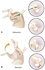
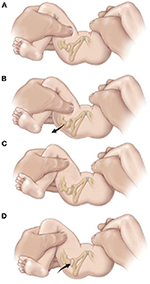
For examining the depth of the acetabulum and joint laxity, the following clinical tests are performed: the Barlow test, the Barden test, the Galeazzi test, and the Ortolani test, all of which were originally adult for employ in humans (1, nine). The Ortolani test is near commonly used in both humans (nine, 51) (Figure 2) and dogs (1, 54) (Figure three). The Barlow test is also commonly used in dogs (1, 54). It should be noted that while on human infants and dogs these tests tin can be directly performed, these tests often require sedation or full general anesthesia when dogs are not cooperative (54) (Figures 2, 3).
Effigy 2. The Barlow and Ortolani test in dogs. (A) "Barlow" (subluxation) test. The dog is positioned in lateral or dorsal recumbency. In lateral recumbency, the examiner is caudal to the dog with one mitt on the distal stifle (flexed to 90 degrees) and the other is dorsal to the pelvis, with the pollex resting over the greater trochanter. The limb is in an adducted position, and force is applied toward the back of the canis familiaris up through the femur (green arrow), causing dorsal subluxation in a hip with articulation laxity. (B) Ortolani (reduction) test. The limb is slowly abducted (yellow pointer) while strength along the axis of the femur is maintained. A positive Ortolani sign is felt when a click or clunk is heard or palpated as the subluxated femoral caput reduces into the acetabulum (ruddy arrow). Figure reproduced without modification from (56) under the Artistic Commons Attribution four.0 International License ().
Effigy 3. The Barlow and Ortolani test in Humans. The Barlow test for developmental dislocation of the hip in a neonate. (A) With the baby supine, the examiner holds both of the child'south knees and gently adducts one hip and pushes posteriorly. (B) When the examination is positive, the examiner will experience the femoral caput make a pocket-size jump (arrow) out of the acetabulum (Barlow's sign). When the force per unit area is released, the head is felt to sideslip dorsum into identify The Ortolani examination for developmental dislocation of the hip in a neonate. (C) The examiner holds the babe's knees and gently abducts the hip while lifting upward on the greater trochanter with two fingers. (D) When the test is positive, the dislocated femoral head will autumn dorsum into the acetabulum (arrow) with a palpable (but not aural) "clunk" every bit the hip is abducted. [Reprinted with permission from Tachdjian's Pediatric Orthopedics (57), Elsevier Publishing].
Since DDH and CHD develop at different rates, a positive event has slightly unlike implications. In humans a positive result indicates subluxation or dislocation of the femoral head typically due to decreased coverage (9, 51). A positive Ortolani test in immature dogs usually points to joint laxity (1, 54) which is a sign of HD in development (58).
Imaging
Radiography is the aureate standard for diagnosing Hard disk drive in dogs (one). Historically, pelvic and hip radiography has been used for diagnosing HD in humans (9, 59). However, in some parts of the worlds Ten-rays have been partially replaced by ultrasound imaging for young patients (60) as classification of HD on Ten-rays is currently considered less reliable earlier ossification of the femoral head center occurs at four–half dozen months (nine, 39). Although at that place are widely accustomed ultrasound classifications, ultrasound images still has drawbacks, such as: high variability and low agreement (61). In dogs the ossification starts at eight weeks, which makes ultrasound less useful as the ossification distorts the view of the acetabulum on ultrasound (54). The manner radiographs are attained and measured is remarkably alike, both in dogs and humans the radiographs are taken in ventrodorsal and anterior-posterior position to mensurate the heart-border (CE)-angle and the Norberg angle (Figure four)(59, 62–64). Also the CE-angle and the Norberg bending, other radiographic parameters can be measured to increment the validity of the diagnosis. However the CE-angle is the most renown (65).
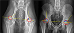
Figure 4. Radiographic diagnostics. Left: Norberg-Angle, take the center of each femoral head (hip brawl) and draw a line between them. Then accept the eye of the femoral head and depict a line to the outer point of the pelvis. The bending between these lines is the Norberg angle. The Norberg angle is calculated for each hip articulation. A normal Norberg Angle ranges from 100/105 to 115 degrees. <100 degrees is dysplastic. Right: CE-angle, take the eye of each femoral head (hip brawl) and depict a line betwixt them. And so take the eye of the femoral head and draw a line to the outer betoken of the pelvis. The bending betwixt these lines is the CE angle. The CE-angle is calculated for each hip joint. A normal CE Angle ranges from 110/115 to 130 degrees. <110 degrees is dysplastic.
Treatment
The available treatments for Hard disk in humans and dogs change when patients develop toward skeletal maturity (39). Young patients accept soft and pliable bone with adept remodeling capabilities due to growth. Therefore, HD treatment tin can focus on stimulating growth past redirecting the femoral head to the center of the triradiate growth plate of the acetabulum in order to create a stable well-covered hip joint (9). As the patient matures the growth potential of bone decreases and the ability to correct the joint relationships with it. When forming a congruent well-covered articulation is no longer an option, osteoarthritis might develop.
Skeletal maturity is reached around 15–xviii years in humans (43) and 1–1.5 years in dogs (66). The triradiate cartilage (acetabular growth plate) closes effectually 14 years of age in humans (67) and around 6 months of age in dogs. At that place is no conspicuously defined separation between treatment options for certain ages and stages of bone development. Therefore, in order to accommodate this review a separation is fabricated between "early" and "tardily" treatment.
Early Non-surgical Treatment
Early on handling of hip dysplasia in humans distinguishes between a (sub-)luxated and a non-luxated hip. A luxated hip needs repositioning first before the acetabular dysplasia tin be treated.
When a dysplastic hip is diagnosed with (sub)luxation of the femoral head, a Pavlik harness is most often applied as first handling. The Pavlik harness uses several straps to flex the hips and knees and prevent adduction, while motility is still possible (9, 68). In a child treated within the 1 weeks after nascency this position forces the femoral head into the acetabular socket and onto the triradiate cartilage. After cosmos of a stable joint, the harness is still worn for 23 h per twenty-four hours until a morphologically normal hip joint is constitute on imaging (39, 68). When a stable reduction of the hip is not reached within 3–four weeks, reposition of the hip under full relaxation nether anesthesia might be tried, with or without adductor tenotomy, followed past a plaster cast usually for three months (9, 68). If repositioning of the hip fails under anesthesia, open reposition of the hip should follow, usually afterwards the age of half-dozen–nine months (9). There is no consensus about optimal treatment length (9, 69). The Pavlik harness has not been described for dogs, since they do not easily accept external hip coaptation devices. Notwithstanding, a somewhat similar concept was used in puppies with genetic predisposition for CHD that were raised in a small cage (1 miii) until they finished growing. This caused them to sit more than frequently with their hind limbs spread (flexion and abduction) and reduced the prevalence of CHD. This method prevents dogs from socializing and is therefore not used in daily practice (48, 54).
In immature dogs with CHD that offset to show clinical signs, usually from historic period 4 to v months, the non-surgical treatment measures are similar to those at older age and therefore will be discussed in more detail in section Late Non-surgical Handling.
Early Surgical Treatment
If non-surgical treatments are ineffective or the child gets older than ix–18 months, open up reduction of the hip articulation can be performed (nine, 46). Open reduction focuses on reducing the subluxated or dislocated hip and creating a stable hip joint, similar to airtight reduction. Open reduction of the hip is usually combined with capsular reefing and the release of the transverse acetabular ligament, and may be combined with an acetabular or femoral osteotomy in order to create a stable well-centered hip (ix). Later on the open up reduction, the child is treated with a spica bandage to maintain the position of the hips (9). In dogs open reduction for a luxated hip due to severe Hard disk drive is never performed. Hip luxation in young dogs with HD, called the luxoid hip, is usually an indication for early on euthanasia, femoral head and cervix resection or total hip replacement from age seven–9 months.
In the older child with residual hip dysplasia, an acetabuloplasty, eastward.k., the Dega, or Pemberton acetabuloplasty is normally used to meliorate centering and acetabular coverage of the femoral head (lxx, 71). While both procedures are dissimilar, both are curved partial osteotomies of the ilium, with a small-scale (bone) graft placed in the osteotomy. This fractional osteotomy causes a hinging effect in the horizontal line of the triradiate cartilage and will reshape the acetabulum, reducing its diameter, all the same increasing depth (72). The Pemberton acetabuloplasty improves anterior and lateral femoral head coverage, simply not coverage of the posterior femoral head. The Dega acetabuloplasty increases the inductive, lateral, and posterior femoral head coverage (72). Acetabuloplasties give the best results when used on patients ii–8 years onetime (46).
Another mutual technique for pelvic osteotomies in young children is the Salter osteotomy (9, 72). This technique is based on a consummate osteotomy of the ilium bone but superior of the acetabulum and redirection of the existing acetabulum (73, 74). Therefore, the Salter osteotomy does non change the shape of the acetabulum. A possible complication described in the Salter osteotomy is instability (71, 74) and another complication for the Salter and Pemberton acetabuloplasty (71) is overcorrection, leading to excessive coverage of the femoral head resulting in femoral acetabular impingement (71, 74).
The majority of early surgical treatments, like the Pemberton and Salter osteotomy used in humans are not applicative in dogs but because CHD is not detected early on plenty in the dog's life. The only comparable treatment in dogs is juvenile pubic symphysiodesis (JPS). The JPS is an early surgical treatment for CHD, and to our knowledge has non been used in humans. JPS is a relatively uncomplicated surgery in which the cartilage of the pubic symphysis is destroyed through electrocauterization. The rut causes the chondrocytes to become necrotic, resulting in premature closure of the pubic symphysis. Since other parts of the pelvis continue to grow, the acetabulum is rotated ventrolateral, similarly to the human Pemberton and Salter osteotomy, which allows for greater femoral head coverage (12, 54, 66). To be effective, JPS should exist performed earlier week xviii in minor dogs or calendar week 22 in large breed dogs (12, 66).
Osteotomies of the femur are often used in humans and infrequently in dogs. In dogs anile ½−ii years the intertrochanteric femoral osteotomy is used to reduce the neck shaft bending (varisation) and anteversion bending, which are often increased in dysplastic hips. The femoral head is moved more medially (12, 75, 76) which helps redirect the femoral caput into the acetabulum (75, 76). This is achieved by removing a bone wedge from the proximal femur and the bone is then stabilized by a claw plate (12, 75, 76). In humans, a femoral osteotomy can be performed sub- or intertrochanteric. The osteotomy also aims to reduce the anteversion (as well called derotational osteotomy) and neck-shaft angle. Femoral osteotomies in humans are oft combined with open up reduction and acetabular osteotomies, between the ages of two–14 years (69).
Late Non-surgical Treatment
There are diverse late non-surgical treatments for dogs and humans with hip dysplasia. To decrease pain (i, 77), reduce lameness (14), and delay onset of osteoarthrosis (i, 66) a variety of treatments are available including medication like NSAIDs (1), reducing torso weight (34, 77), life style changes including training of pelvic muscles, practice programs and the limiting sudden explosive movements (like throwing a brawl for dogs). On average, weight loss in dogs delays surgery for another iii years (10) and in overweight dogs and humans x% body weight reduction is associated with a relieve in symptoms and signs (xiv, 77). Another non-surgical intervention is the nutraceutical market, which is especially big in the veterinary market. Nutraceuticals are nutrient additives or supplements that are purported to have a disease modifying potential in hip dysplasia and osteoarthritis, merely likewise other conditions. An case of a nutraceutical is Polysulfanated glycosaminoglycans (PSGAGs) which proposedly stimulates collagen synthesis and inhibits the breakdown of collagen (13) which may help reduce subluxation (54).
Another pick for early on not-surgical handling is physiotherapy (78, 79). In both dogs and humans physiotherapy and hydrotherapy is an important component get-go as a conservative treatment option only also equally an important attribute in post-surgical rehabilitation (78, 79).
Late Surgical Handling
Originally designed for humans with Hard disk, triple pelvic osteotomy (TPO) has also go a successful procedure for dogs with HD (12, 80)(Figure five). This surgery can be used in young dogs (one, 54, 81), but more than frequently in adolescents (46, 82, 83) and young adults (82, 83) without or with minimal degenerative joint damage (one, 49, 54, 83). In dogs, the surgery is preferably performed before full skeletal maturity is reached, while in humans information technology can be used both earlier and after the triradiate cartilage closes (9, 83). However, humans have more early surgical treatments available (e.grand., Salter & Pemberton), deferring the more invasive TPO to older patients. Over the years there accept been many changes in specific surgical techniques, just the general outline of TPO remains the same. Osteotomies are made in the pubic, ischial, and iliac bones, and the acetabulum is subsequently rotated ventrally to improve femoral head coverage and increase the load bearing area (eighty) (Figure 5). The acetabulum is then fixated in place by plates, screws, or K-wire. Clinical reduction of lameness after TPO, and comeback in weight bearing of 86–92% is reported in dogs (75, 76, 81). The joint laxity is reduced post-obit TPO in dogs (lxxx, 81), only degenerative changes cannot exist stopped completely (81). In humans, TPO causes a long term reduction of pain and improvement of part (84) and a (total hip free) survival of 68% later on 25 years is reported. Recently, dual pelvic osteotomy (DPO) has been recommended in dogs (75, 85), it has also been described in humans (72). DPO is similar to TPO, with a faster mail service-operative recovery, every bit in that location is no osteotomy of the ischium and therefore no pelvic discontinuity (75, 85).
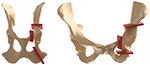
Figure 5. Canine left and human being correct triple pelvic osteotomies (purple planes) are made in the pubic, ischial and iliac bones and after the acetabulum is rotated ventrally (dogs) or anteriorly (humans) to improve femoral head coverage and increment the load bearing area.
Shelf arthroplasty is a commonly used salvage procedure for Hd in humans (86). It involves the placement of an autologous bone graft outside of the joint capsule superior to the acetabulum (87, 88). The graft can be impacted into the os or be held in identify past a spiral (88), improving the support structure of the joint (9). Capsular metaplasia causes the improvement of the articulating surface. The improved support and improved femoral caput coverage helps ameliorate the weight begetting surface (9) and delays the progression of OA. This procedure is preferably performed in younger patients with minimal arthritic changes, however information technology is generally reserved as a salvage process equally other treatments are not eligible. The survival of the shelf process can exist up to 72% at 35 years of follow-up (86). A like procedure chosen the biocompatible osteoconductive polymer (BOP) procedure has been described every bit an alternative to TPO in boyish dogs (12, 75, 76). Instead of autologous os graft, biocompatible osteoconductive fibers were used to increment coverage, because the fibers were expected to promote bony ingrowth (12, 75, 76). Despite that the shelf procedure was successful in humans, BOP in dogs never became a common procedure because of uncontrolled bone growth (75, 76). New procedures involving 3D-printed titanium shelfs (89) or 3D-printed biodegradable magnesium phosphate shelfs (ninety) are notwithstanding beingness developed in dogs and when successful these procedures hopefully find their fashion back to the homo dispensary.
While being i of the most effective procedures, total hip arthroplasty (THA) in humans and dogs (12) is oftentimes postponed as the concluding treatment choice (Figure half dozen). When young patients with a demanding lifestyle receive a THA they may need one or more than revisions in their lifetime due to implant clothing. Nonetheless, every revision is more difficult to perform due to fibrosis in the perioperative area. Therefore, in humans, the demand for THA is preferably postponed across the age of 60 to prevent revisions in the long term. Although THA has been available for dogs for three decades, it remains an expensive handling option, especially when the owners have no insurance (49). Likewise, THA in humans can get technically enervating due to anatomical differences in dysplastic hips making it difficult to ream a large enough bony bed to back up an acetabular cup (91). In dogs the process is technically enervating due to breed anatomic differences simply can be used in dogs of any size or shape when aged ix months or older (12).
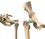
Figure half dozen. The comparable gear up-up of the total hip arthroplasty in humans and dogs. On the left the set-upwardly in dogs and on the right the set-up in humans.
It is proficient to note that implant improvements accept benefited for cross species research. For example, due to the active nature of dogs, the THA materials demand is loftier and companies specializing in canine THA have benefited from the prosthetic cognition beingness researched and developed for human medicine. For example, similar durable materials developed for human cups and stems are translated to the dog THA assuasive dogs to perform without the need for revision beyond a decade lifetime, with a biomechanically enervating lifestyle asking for more cyclic loading of their implants than humans. Vice versa, in dogs new products are adult e.g., to decrease stem loosening, because dogs demand firsthand total weightbearing after surgery due to there not-compliance to life style restrictions. One example of a successful concept in THA surgery in dogs (Zürich cementless THA) is the immediate stem screw fixation at the medial femoral cortex instead of printing fit fixation (92). Likewise, in a few years more developments might be translated dorsum from the veterinary to the homo marketplace.
One of the least performed in humans but most commonly executed salvage procedures in dogs is the femoral head and neck excision (75, 76). The surgery is relatively piece of cake to perform and has low costs, and is most effective in dogs with low torso weight (75, 93). The removal of the femoral head and neck leads to a fibrotic pseudoarthrosis (false joint), allowing for relative pain gratis movement (12, 49). Femoral head and neck excision mostly has good results in relieving hurting, however possible side effects are extensive rehabilitation, decreased range of motion, muscle atrophy and limping due to decreased limb length (12, 75, 76, 93). Important factors in the issue are body size (12, 75, 76), dog temperament and activity (12, 94). This procedure performed in humans is chosen a "Girdle rock" procedure, but only every bit the terminal option (94).
Conclusion
In this review we described the anatomy, etiology, development, diagnostics and handling of Hard disk in humans and dogs. Humans and dogs take like anatomy on micro- and macroscopic levels. Hd every bit an orthopedic condition has many overlying characteristics in humans and dogs in terms of etiology and pathogenesis. As well, treatment of Hd shows many similarities. In that location is much parallel employ of early on and after growth (conservative) treatments and interventions. Moreover, many of the surgical treatments for HD that were adult for humans accept first been tested in experimental dogs. Procedures that became successful in humans constitute their way to the veterinary field and are at present usually used in companion animal clinics. We suggest that further exchange betwixt research on Hard disk in humans and dogs can be benign for the treatment of HD in humans and dogs.
Author Contributions
BW, BM, RS, HW, and KW: conceptualized the report. KW, MM, and NH: nerveless the information. KW, MT, MM, NH, and RS: analyzed the information. HW, BM, MT, and BW: supervised the projection. KW, MT, RS, NH, MM, HW, BM, and BW: wrote, edited and reviewed the newspaper. All authors have read and canonical the terminal submitted manuscript.
Funding
KW and HW report a Governmental inquiry grant from Interreg VA Flanders—Kingdom of the netherlands program, during the conduct of the study; KW and HW report grants from the Smart industry project 15479 funded through governmental grant by NWO domain TTW, during the conduct of the written report; MT, HW, and BM study funding from the Dutch Arthritis Society (LLP12/22). The funding sources played no role in the study pattern, information analysis and interpretation, nor drafting of the manuscript or the conclusion to submit it for publication.
Conflict of Interest
The authors declare that the research was conducted in the absence of any commercial or financial relationships that could be construed as a potential conflict of interest.
Publisher's Note
All claims expressed in this article are solely those of the authors and do not necessarily represent those of their affiliated organizations, or those of the publisher, the editors and the reviewers. Whatsoever production that may exist evaluated in this article, or claim that may be made by its manufacturer, is not guaranteed or endorsed past the publisher.
References
2. Olmstead ML, Linda LD. Small Animal Orthopedics. St. Louis: Mosby (1995). p. 365–8.
Google Scholar
three. Stein-Zamir C, Volovik I, Rishpon South, Sabi R. Developmental dysplasia of the hip: risk markers, clinical screening and outcome. Pediatr Int. (2008) 50:341–5. doi: ten.1111/j.1442-200X.2008.02575.ten
PubMed Abstract | CrossRef Total Text | Google Scholar
5. Vanden Berg-Foels WS, Todhunter RJ, Schwager SJ, Reeves AP. Event of early postnatal body weight on femoral head ossification onset and hip osteoarthritis in a canine model of developmental dysplasia of the hip. Pediatr Res. (2006) sixty:549–54. doi: 10.1203/01.pdr.0000243546.97830.a0
PubMed Abstract | CrossRef Total Text | Google Scholar
vi. Roberts T, McGreevy PD. Option for brood-specific long-bodied phenotypes is associated with increased expression of canine hip dysplasia. Vet J. (2010) 183:266–72. doi: 10.1016/j.tvjl.2009.11.005
PubMed Abstract | CrossRef Full Text | Google Scholar
7. Worth AJ, Bridges JP, Cave NJ, Jones G. Seasonal variation in the hip score of dogs as assessed past the New Zealand Veterinarian Association hip dysplasia scheme. N Z Vet J. (2012) 60:110–4. doi: x.1080/00480169.2011.636730
PubMed Abstract | CrossRef Full Text | Google Scholar
8. Rettenmaier JL, Keller GG, Lattimer JC, Corley EA, Ellersieck MR. Prevalence of canine hip dysplasia in a veterinary instruction hospital population. Vet Radiol Ultrasound. (2002) 43:313–viii. doi: 10.1111/j.1740-8261.2002.tb01010.x
PubMed Abstract | CrossRef Full Text | Google Scholar
ix. Kotlarsky P, Haber R, Bialik V, Eidelman M. Developmental dysplasia of the hip: what has changed in the concluding 20 years? Earth J Orthop. (2015) half dozen:886–901. doi: 10.5312/wjo.v6.i11.886
PubMed Abstract | CrossRef Full Text | Google Scholar
10. Smith GK, Karbe GT, Agnello KA, McDonald-Lynch MB. Pathogenesis, diagnosis and control of canine hip dysplasia. In: Veterinarian Surgery: Pocket-sized Brute. 1st ed. St. Louis: Elsevier (2012). p. 824–48.
PubMed Abstract
11. Barr ARS, Denny HR, Gibbs DC. Clinical hip dysplasia in growing dogs : the long-term results of bourgeois management. J Small Anim Pract. (1987) 28:243–52. doi: ten.1111/j.1748-5827.1987.tb03879.x
CrossRef Full Text | Google Scholar
14. Impellizeri JA, Tetrick MA, Muir P. Outcome of weight reduction on clinical signs of lameness in dogs with hip osteoarthritis. J Am Vet Med Assoc. (2000) 216:1089–91. doi: x.2460/javma.2000.216.1089
PubMed Abstract | CrossRef Total Text | Google Scholar
15. Bonneau North, Baylac Thou, Gagey O, Tardieu C. Functional integrative analysis of the man hip joint: the three-dimensional orientation of the acetabulum and its relation with the orientation of the femoral cervix. J Hum Evol. (2014) 69:55–69. doi: ten.1016/j.jhevol.2013.12.013
PubMed Abstruse | CrossRef Total Text | Google Scholar
18. Gruss LT, Schmitt D. The evolution of the human pelvis: changing adaptations to bipedalism, obstetrics and thermoregulation. Philos Trans R Soc Lond B Biol Sci. (2015) 370:20140063. doi: 10.1098/rstb.2014.0063
PubMed Abstract | CrossRef Full Text | Google Scholar
twenty. Soontornvipart Yard, Chalayon P, Tangwongsan C. Continuing analysis of healthy and abnormal canines using forcefulness platform system. In: 2013 tenth International Conference on Electrical Applied science/Electronics, Computer, Telecommunications and It. IEEE (2013) p. 1–six.
Google Scholar
22. Nunamaker DM, Blauner PD. Normal and Abnormal Gait. Textb Small Anim Orthop. Ithaca, NY: International Veterinarian Data Services (IVIS) (1985).
Google Scholar
23. Byrne DP, Mulhall KJ, Bakery JF. Anatomy & Biomechanics of the Hip. Open Sport Med J. (2010) (4):51–vii. doi: x.2174/1874387001004010051
CrossRef Total Text | Google Scholar
24. Pearce AI, Richards RG, Milz South, Schneider Due east, Pearce SG. Animal models for implant biomaterial inquiry in bone: a review. Eur Prison cell Mater. (2007) thirteen:1–10. doi: 10.22203/eCM.v013a01
PubMed Abstract | CrossRef Full Text | Google Scholar
25. Eitel F, Klapp F, Jacobson W, Schweiberer L. Bone regeneration in animals and in man. A contribution to understanding the relative value of brute experiments to human pathophysiology. Arch Orthop Trauma Surgery. (1981) 99:59–64. doi: 10.1007/BF00400911
PubMed Abstract | CrossRef Total Text | Google Scholar
26. Rhinelander FW, Nelson CL, Stewart RD, Stewart CL. Experimental reaming of the proximal femur and acrylic cement implantation: vascular and histologic effects. Clin Orthop Relat Res. (1979) (141):74–89. doi: 10.1097/00003086-197906000-00009
PubMed Abstract | CrossRef Full Text | Google Scholar
27. Goel VK, Drinker H, Panjabi MM, Strongwater A. Option of an animal model for implant fixation studies: anatomical aspects. Yale J Biol Med. (1982) 55:113–22.
PubMed Abstract | Google Scholar
28. Aerssens J, Boonen S, Lowet G, Dequeker J. Interspecies differences in bone composition, density, and quality: potential implications for in vivo bone research. Endocrinology. (1998) 139:663–70. doi: 10.1210/endo.139.ii.5751
PubMed Abstract | CrossRef Total Text | Google Scholar
29. Ibrahim T, Riaz K, Hegazy A. The prevalence of developmental dysplasia of the hip in idiopathic clubfoot: a systematic review and meta-assay. Int Orthop. (2015) 39:1371–eight. doi: x.1007/s00264-015-2757-z
PubMed Abstruse | CrossRef Total Text | Google Scholar
31. Sugano Northward, Noble PC, Kamaric East, Salama JK, Ochi T, Tullos HS. The morphology of the femur in developmental dysplasia of the hip. J Bone Articulation Surg Br. (1998) 80:711–nine. doi: x.1302/0301-620X.80B4.0800711
PubMed Abstract | CrossRef Full Text | Google Scholar
32. Krontveit RI, Nødtvedt A, Sævik BK, Ropstad Eastward, Skogmo HK, Trangerud C, et al. Prospective study on canine hip dysplasia and growth in a cohort of four large breeds in Norway (1998-2001). Prev Vet Med. (2010) 97:252–63. doi: ten.1016/j.prevetmed.2010.09.015
PubMed Abstract | CrossRef Full Text | Google Scholar
33. Kealy RD, Olsson SE, Monti KL, Lawler DF, Biery DN, Helms RW, et al. Effects of limited food consumption on the incidence of hip dysplasia in growing dogs. Journal-American Vet Med Assoc. (1992) 201:857.
PubMed Abstract | Google Scholar
34. Karlson EW, Mandl LA, Aweh GN, Sangha O, Liang MH, Grodstein F. Total hip replacement due to osteoarthritis: the importance of age, obesity, and other modifiable risk factors. Am J Med. (2003) 114:93–8. doi: 10.1016/S0002-9343(02)01447-X
PubMed Abstract | CrossRef Full Text | Google Scholar
35. Smith GK, Paster ER, Powers MY, Lawler DF, Biery DN, Shofer FS, et al. Lifelong diet restriction and radiographic evidence of osteoarthritis of the hip joint in dogs. J Am Vet Med Assoc. (2006) 229:690–3. doi: 10.2460/javma.229.v.690
PubMed Abstract | CrossRef Full Text | Google Scholar
36. Atalar H, Gunay C, Yavuz OY, Camurdan Advertizement, Uras I, Eren A. Maternal summit and infant body mass index are possible risk factors for developmental dysplasia of the hip in female infants. Acta Med Okayama. (2015) 69:349–51.
PubMed Abstract | Google Scholar
37. Steinetz BG, Williams AJ, Lust M, Schwabe C, Büllesbach EE, Goldsmith LT. Transmission of relaxin and estrogens to suckling canine pups via milk and possible clan with hip articulation laxity. Am J Vet Res. (2008) 69:59–67. doi: 10.2460/ajvr.69.i.59
PubMed Abstract | CrossRef Full Text | Google Scholar
38. Flückiger MA, Steffen F, Hässig M, Morgan JP. Asymmetrical lumbosacral transitional vertebrae in dogs may promote asymmetrical hip articulation evolution. Vet Comp Orthop Traumatol. (2017) 30:137–42. doi: ten.3415/VCOT-16-05-0072
PubMed Abstract | CrossRef Full Text | Google Scholar
39. Schwend RM, Shaw BA, Segal LS. Evaluation and treatment of developmental hip dysplasia in the newborn and baby. Pediatr Clin Due north Am. (2014) 61:1095–107. doi: x.1016/j.pcl.2014.08.008
PubMed Abstract | CrossRef Full Text | Google Scholar
xl. Kutlu A, Memik R, Mutlu M, Kutlu R, Arslan A. Congenital dislocation of the hip and its relation to swaddling used in Turkey. J Pediatr Orthop. (1992) 12:598–602. doi: 10.1097/01241398-199209000-00006
PubMed Abstruse | CrossRef Full Text | Google Scholar
41. De Pellegrin M, Moharamzadeh D. Developmental dysplasia of the hip in twins: the importance of mechanical factors in the etiology of DDH. J Pediatr Orthop. (2010) 30:774–8. doi: ten.1097/BPO.0b013e3181fc35c0
PubMed Abstract | CrossRef Full Text | Google Scholar
42. Manoukian D, Rehm A. Oligohydramnios: Should information technology be considered a gamble factor for developmental dysplasia of the hip? J Pediatr Orthop Part B. (2019) 28:442–5. doi: x.1097/BPB.0000000000000624
PubMed Abstract | CrossRef Full Text | Google Scholar
43. Ford CA, Nowlan NC, Thomopoulos S, Killian ML. Furnishings of imbalanced muscle loading on hip joint development and maturation. J Orthop Res. (2016) 35:1128–36 doi: 10.1002/jor.23361
PubMed Abstract | CrossRef Full Text | Google Scholar
45. Sakkers R, Pollet V. The natural history of abnormal ultrasound findings in hips of infants under vi months of age. J Child Orthop. (2018) 12:302–7. doi: 10.1302/1863-2548.12.180056
PubMed Abstract | CrossRef Full Text | Google Scholar
47. Tobias KM, Johnston SA. Veterinary surgery: small beast-E-Book: 2-book set. Elsevier Health Sciences. (2013).
Google Scholar
49. Rawson EA, Aronsohn MG, Burk RL. Simultaneous bilateral femoral head and cervix ostectomy for the treatment of canine hip dysplasia. J Am Anim Hosp Assoc. (2005) 41:166–70. doi: 10.5326/0410166
PubMed Abstruse | CrossRef Total Text | Google Scholar
50. Gatineau Thousand, Dupuis J, Beauregard 1000, Charette B, Breton Fifty, Beauchamp 1000, et al. Palpation and dorsal acetabular rim radiographic projection for early detection of canine hip dysplasia: a prospective study. Vet Surg. (2012) 41:42–53. doi: ten.1111/j.1532-950X.2011.00926.ten
PubMed Abstract | CrossRef Full Text | Google Scholar
51. Shorter D, Hong T, Osborn DA. Screening programmes for developmental dysplasia of the hip in newborn infants. Cochrane database Syst Rev. (2011) (9):CD004595. doi: 10.1002/14651858.CD004595.pub2
PubMed Abstract | CrossRef Full Text | Google Scholar
52. Kang MS, Han GW, Kam Thousand, Park S-S. Clinical significance of disproportionate skin folds in the medial thigh for the infantile screening of developmental dysplasia of the hip. Pediatr Neonatol. (2019) lx:570–six. doi: x.1016/j.pedneo.2019.02.004
PubMed Abstract | CrossRef Total Text | Google Scholar
54. Ginja MMD, Silvestre AM, Gonzalo-Orden JM, Ferreira AJA. Diagnosis, genetic command and preventive management of canine hip dysplasia: a review. Vet J. (2010) 184:269–76. doi: ten.1016/j.tvjl.2009.04.009
PubMed Abstract | CrossRef Full Text | Google Scholar
55. Hielm-Björkman AK, Kuusela Eastward, Liman A, Markkola A, Saarto E, Huttunen P, et al. Evaluation of methods for assessment of pain associated with chronic osteoarthritis in dogs. J Am Vet Med Assoc. (2003) 222:1552–eight. doi: x.2460/javma.2003.222.1552
PubMed Abstract | CrossRef Full Text | Google Scholar
56. Kyriazis A, Prassinos NN. Canine hip dysplasia: office i: aetiopathogenesis & diagnostic arroyo. Hell J Companion Anim Med. (2016) 5:22–47.
Google Scholar
57. Herring JA. Tachdjian's Pediatric Orthopaedics: From the Texas Scottish Rite Hospital for Children East-Volume. Philadelphia, PA: Elsevier (2020).
Google Scholar
58. Ginja MMD, Ferreira AJ, Jesus SS, Melo-Pinto P, Bulas-Cruz J, Orden MA, et al. Comparison of clinical, radiographic, computed tomographic, and magnetic resonance imaging methods for early prediction of canine hip laxity and dysplasia. Vet Radiol Ultrasound. (2009) 50:135–43. doi: 10.1111/j.1740-8261.2009.01506.x
PubMed Abstract | CrossRef Full Text | Google Scholar
59. Wiberg M. Studies on dysplastic acetabula and congenital subluxation of the hip joint: with special reference to the complication of osteoarthritis. Acta Chir Scand. (1939) 83:53–68.
PubMed Abstruse | Google Scholar
60. Storer SK, Skaggs DL. Developmental dysplasia of the hip. Am Fam Medico. (2006) 74:1310–6.
Google Scholar
61. Quader N, Schaeffer EK, Hodgson AJ, Abugharbieh R, Mulpuri Thou A. systematic review and meta-analysis on the reproducibility of ultrasound-based metrics for assessing developmental dysplasia of the hip. J Pediatr Orthop. (2018) 38:e305–xi. doi: 10.1097/BPO.0000000000001179
PubMed Abstruse | CrossRef Full Text | Google Scholar
62. Spud SB, Ganz R, Müller ME. The prognosis in untreated dysplasia of the hip. A study of radiographic factors that predict the outcome. J Bone Jt Surg Am. (1995) 77:985–nine. doi: 10.2106/00004623-199507000-00002
PubMed Abstract | CrossRef Full Text | Google Scholar
63. Murphy SB, kijewski PK, Millis MB, Harless A. Acetabular dysplasia in the adolescent and young adult. Clin Orthop Relat Res. (1990) 261:214–23. doi: 10.1097/00003086-199012000-00023
PubMed Abstract | CrossRef Total Text | Google Scholar
64. Werner CML, Ramseier LE, Ruckstuhl T, Stromberg J, Copeland CE, Turen CH, et al. Normal values of Wiberg's lateral center-edge bending and Lequesne's acetabular alphabetize–a coxometric update. Skeletal Radiol. (2012) 41:1273–8. doi: 10.1007/s00256-012-1420-7
PubMed Abstract | CrossRef Full Text | Google Scholar
65. Monazzam Due south, Bomar JD, Cidambi K, Kruk P, Hosalkar H. Lateral center-border bending on conventional radiography and computed tomography. Clin Orthop Relat Res. (2013) 471:2233–7. doi: 10.1007/s11999-012-2651-half-dozen
PubMed Abstract | CrossRef Total Text | Google Scholar
66. Vezzoni A, Dravelli G, Vezzoni L, De Lorenzi G, Corbari A, Cirla A, et al. Comparison of conservative management and juvenile pubic symphysiodesis in the early treatment of canine hip dysplasia. Vet Comp Orthop Traumatol. (2008) 21:267–79. doi: 10.1055/southward-0037-1617372
PubMed Abstract | CrossRef Total Text | Google Scholar
67. Parvaresh KC, Pennock AT, Bomar JD, Wenger DR, Upasani VV. Analysis of acetabular ossification from the triradiate cartilage and secondary centers. J Pediatr Orthop. (2018) 38:e145–50. doi: 10.1097/BPO.0000000000001120
PubMed Abstract | CrossRef Full Text | Google Scholar
seventy. López-Carreño E, Carillo H, Gutiérrez 1000. Dega vs. Salter osteotomy for the treatment of developmental dysplasia of the hip. J Pediatr Orthop B. (2008) 17:213–21. doi: 10.1097/BPB.0b013e32830850eb
PubMed Abstruse | CrossRef Full Text | Google Scholar
71. Castañeda P, Vidal-Ruiz C, Méndez A, Salazar DP, Torres A. How ofttimes does femoroacetabular impingement occur afterward an innominate osteotomy for acetabular dysplasia? Clin Orthop Relat Res. (2016) 474:1209–15. doi: 10.1007/s11999-016-4721-7
PubMed Abstract | CrossRef Full Text | Google Scholar
72. Sales de. Gauzy J. Pelvic reorientation osteotomies and acetabuloplasties in children Surgical technique. Orthop Traumatol Surg Res. (2010) 96:793–nine. doi: 10.1016/j.otsr.2010.07.004
PubMed Abstract | CrossRef Total Text | Google Scholar
73. Wang CW, Wang TM, Wu KW, Huang SC, Kuo KN. The comparative, long-term effect of the salter osteotomy and pemberton acetabuloplasty on pelvic height, scoliosis and functional outcome. Bone Joint J. (2016) 98:1145–l. doi: 10.1302/0301-620X.98B8.37215
PubMed Abstract | CrossRef Total Text | Google Scholar
74. Wang C-W, Wu K-W, Wang T-M, Huang S-C, Kuo KN. Comparison of acetabular anterior coverage afterwards salter osteotomy and pemberton acetabuloplasty: a long-term followup. Clin Orthop Relat Res. (2014) 472:1001–nine. doi: x.1007/s11999-013-3319-6
PubMed Abstract | CrossRef Full Text | Google Scholar
75. Raghuvir HB, Shivrajsinh KJ, Dipak NS, Harit DB, Chirag AB, Naresh HK. Treatment of canine hip dysplasia: a review. J Anim Sci Adv. (2013) iii:589–97.
PubMed Abstruse | Google Scholar
76. Remedios AM, Chips CL. Treatment of canine hip dysplasia: a review. Can Vet J = La Rev vétérinaire Can. (1995) 36:503–9.
PubMed Abstract | Google Scholar
77. Bliddal H, Leeds AR, Christensen R. Osteoarthritis, obesity and weight loss: testify, hypotheses and horizons - a scoping review. Obes Rev. (2014) 15:578–86. doi: 10.1111/obr.12173
PubMed Abstruse | CrossRef Full Text | Google Scholar
78. Bockstahler B, Levine D, Maierl J, Millis D, Wittek K. Essential Facts of Physical Medicine, Rehabilitation and Sports Medicine in Companion Animals. Babenhausen: VBS GmbH (2019).
79. Retchford Th, Crossley KM, Grimaldi A, Kemp JL, Cowan SM. Can local muscles broaden stability in the hip? A narrative literature review. J Musculoskelet Neuronal Interact. (2013) xiii:1−12.
PubMed Abstract | Google Scholar
80. Hara Y, Harada Y, Fujita Y, Taoda T, Nezu Y, Yamaguchi Due south, et al. Changes of hip articulation congruity after triple pelvic osteotomy in the dog with hip dysplasia. J Vet Med Sci. (2002) 64:933–6. doi: x.1292/jvms.64.933
PubMed Abstract | CrossRef Total Text | Google Scholar
81. McLaughlin RM, Miller CW, Taves CL, Hearn TC, Palmer NC, Anderson GI. Force plate assay of triple pelvic osteotomy for the handling of canine hip dysplasia. Vet Surg. (1991) twenty:291–7. doi: x.1111/j.1532-950X.1991.tb01270.x
PubMed Abstract | CrossRef Full Text | Google Scholar
82. de Kleuver Yard, Kapitein PJ, Kooijman MA, van Limbeek J, Pavlov Pow, Veth RP. Acetabular coverage of the femoral caput after triple pelvic osteotomy: no relation to outcome in 51 hips followed for 8-fifteen years. Acta Orthop Scand. (1999) 70:583–8. doi: 10.3109/17453679908997846
PubMed Abstract | CrossRef Total Text | Google Scholar
83. Mimura T, Mori K, Kawasaki T, Imai Southward, Matsusue Y. Triple pelvic osteotomy: Report of our mid-term results and review of literature. World J Orthop. (2014) five:14–22. doi: 10.5312/wjo.v5.i1.14
PubMed Abstruse | CrossRef Full Text | Google Scholar
84. van Stralen RA, van Hellemondt GG, Ramrattan NN, de Visser E, de Kleuver One thousand. Tin can a triple pelvic osteotomy for developed symptomatic hip dysplasia provide relief of symptoms for 25 years? Clin Orthop Relat Res. (2013) 471:584–90. doi: 10.1007/s11999-012-2701-0
PubMed Abstract | CrossRef Total Text | Google Scholar
85. Vezzoni A, Boiocchi S, Vezzoni L, Vanelli AB, Bronzo V. Double pelvic osteotomy for the treatment of hip dysplasia in young dogs. Vet Comp Orthop Traumatol. (2010) 23:444–52. doi: 10.3415/VCOT-x-03-0034
PubMed Abstract | CrossRef Full Text | Google Scholar
86. Willemsen K, Doelman CJ, Sam ASY, Seevinck PR, Sakkers RJB, Weinans H, et al. Long-term outcomes of the hip shelf arthroplasty in adolescents and adults with rest hip dysplasia: a systematic review. Acta Orthop. (2020) 91:383–9. doi: 10.1080/17453674.2020.1747210
PubMed Abstruse | CrossRef Full Text | Google Scholar
87. Migaud H, Chantelot C, Giraud F, Fontaine C, Duquennoy A. Long-term survivorship of hip shelf arthroplasty and Chiari osteotomy in adults. Clin Orthop Relat Res. (2004) (418):81–six. doi: 10.1097/00003086-200401000-00014
PubMed Abstract | CrossRef Full Text | Google Scholar
88. Bartoníček J, Vávra J, Chochola A. Bosworth hip shelf arthroplasty in developed dysplastic hips: x to twenty three year results. Int Orthop. (2012) 36:2425–31. doi: 10.1007/s00264-012-1665-8
PubMed Abstract | CrossRef Full Text | Google Scholar
89. Willemsen K, Tryfonidou Chiliad, Sakkers R, Castelein RM, Zadpoor AA, Seevinck P, et al. Patient-specific 3D-printed shelf implant for the handling of hip dysplasia: Anatomical and biomechanical outcomes in a canine model. J Orthop Res. (2021). doi: 10.1002/jor.25133
PubMed Abstract | CrossRef Full Text | Google Scholar
90. Golafshan N, Willemsen Thousand, Kadumudi FB, Vorndran East, Dolatshahi-Pirouz A, Weinans H, et al. 3D-printed regenerative magnesium phosphate implant ensures stability and restoration of hip dysplasia. Adv Healthc Mater. x:east 101051. doi: 10.1002/adhm.202101051
PubMed Abstract | CrossRef Full Text | Google Scholar
91. Bicanic G, Barbarian Yard, Bohacek I, Aljinovic A, Delimar D. Electric current concept in dysplastic hip arthroplasty: Techniques for acetabular and femoral reconstruction. World J Orthop. (2014) five:412–24. doi: 10.5312/wjo.v5.i4.412
PubMed Abstract | CrossRef Full Text | Google Scholar
93. Off West, Matis U. Excision arthroplasty of the hip articulation in dogs and cats. clinical, radiographic, and gait assay findings from the department of surgery, veterinary faculty of the ludwig-maximilians-university of munich, federal republic of germany. 1997. Vet Comp Orthop Traumatol. (2010) 23:297–305.
PubMed Abstruse | Google Scholar
94. Olsson SE, Figarola F, Suzuki K. Femoral head excision arthroplasty. A save performance in severe hip dysplasia in dogs. Clin Orthop Relat Res. (1969) 62:104–12. doi: ten.1097/00003086-196901000-00013
PubMed Abstract | CrossRef Full Text | Google Scholar
Source: https://www.frontiersin.org/articles/10.3389/fvets.2021.791434/full
0 Response to "Hip Dysplasia Pain in Dogs Pain Meds Reviews"
Post a Comment